pleural effusion cat ultrasound
To mark the optimal site for drainage and perform the procedure if required. 32 2 Eibenberger formula.

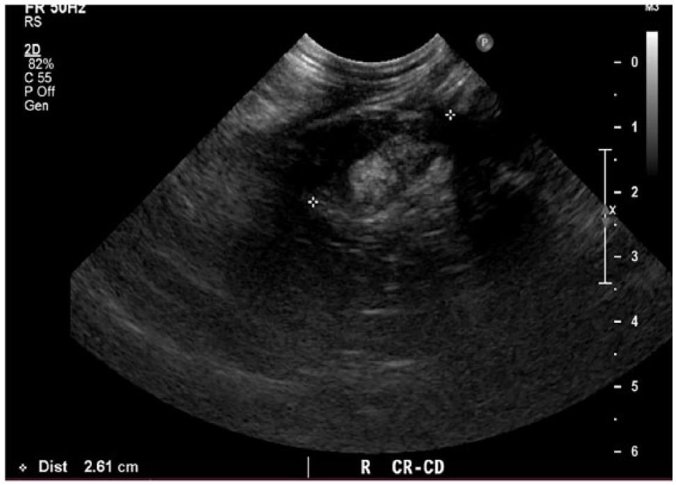
Abdominal Ultrasound Images Of A 5 Year Old Cat Case 1 With An Download Scientific Diagram
The aspect of pleural effusion on ultrasound can suggest the nature of the fluid although a definitive diagnosis requires a thoracentesis in order to allow physical chemical and.

. The success rate is low when the effusion is loculated and septated. In some cases ultrasound may also be. Abdominal ultrasounds were performed in 70 cats with pleural effusion and revealed concurrent abdominal effusion in 59 of these cats.
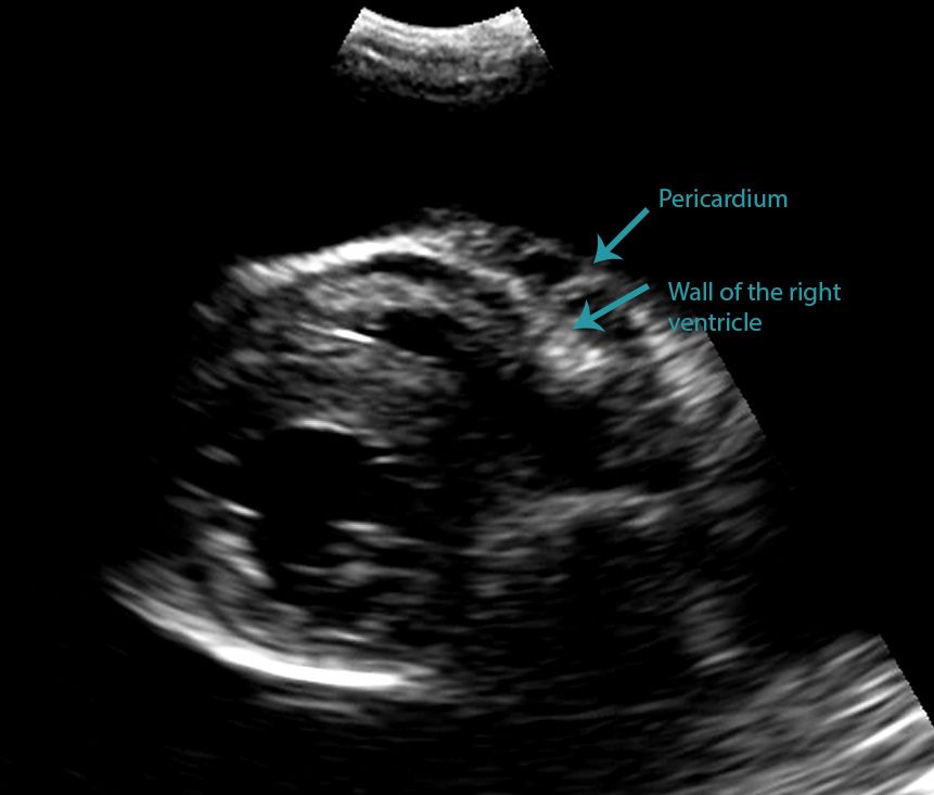
For those who are new to imaging around the heart with ultrasound differentiating a pericardial from a pleural effusion can be tricky particularly when the pleural effusion is circumferential around the heart. Unlike with a pericardial effusion in the case of accumulation of fluid in the pleural space there is no collapse of the heart walls. Using a handheld ultrasound device is a quick way to identify the presence of pleural fluid and therefore can help in diagnosing pleural effusion.
These four signs the diaphragm sign the displaced crus sign the interface sign and the bare area sign are reliable when only one abnormal fluid collection is present. The aims of ultrasound guided assessment of pleural effusion are. Thoracic ultrasound TUS helps clinicians not only to visualize pleural effusion but also to distinguish between the different types.
Measurement of a pleural effusion volume with point-of-care ultrasonography may be a useful tool for intensivists and is an active area of research in critical care 7. The pleural effusion volume was calculated volumetrically from the CT scan data. 2006 Intensive care medicine.
To determine and describe the size and site of the effusion. Many studies have demonstrated the usefulness of ultrasound for the identification and characterization of pleural fluid. 91 Pediatric abdomen and retroperitoneum 92 Pediatric urinary tract 93 Pediatric scrotum 94 Pediatric gynaecological pathology and infant breast 95 Pediatric head and neck 96 Neonatal brain and spine 97 Infant hip and knee 98 Pediatric thorax.
This procedure removes excess fluid from the pleural space using a needle which not only relieves pressure on your cats lungs but also provides your. The therapeutic intervention also provides your first diagnostic test. Pleural effusion PLEFF mostly caused by volume overload congestive heart failure and pleuropulmonary infection is a common condition in critical care patients.
Pleural effusions as small as 35 ml may be detected by ultrasound. As an article in Acta Radiologica identified Due to its ease of use and its high diagnostic yield hand-carried ultrasound systems of the latest generation constitute a helpful technique for. In the following article we present two cases concluding with a third case in which both types of effusion can be seen simultaneously.
Ultrasound estimation of volume of pleural fluid in mechanically ventilated patients. It is much better than a chest X-ray at showing if there is any fluid there and how much there is. The scan involves putting some jelly on the skin over the ribcage and then a probe made of plastic used to scan the fluid.
The trocar technique is faster and easier. Effusion volume mL 476 x maximum perpendicular distance between the pulmonary surface and chest wall at maximal inspiration mm 837. If the FAST ultrasound does reveal pleural effusion thoracentesis can be carried out.
As an article in Acta Radiologica identified Due to its ease of use and its high diagnostic yield hand-carried ultrasound systems of the latest generation constitute a helpful technique for. Ultrasound is a simple scan that is done on the chest to determine how much fluid is present and what the fluid looks like. In the latter situations therapeutic intervention must be initiated quickly to prevent respiratory arrest.
Ultrasound can be used in the assessment of pleural effusion volume. About Press Copyright Contact us Creators Advertise Developers Terms Privacy Policy Safety How YouTube works Test new features Press Copyright Contact us Creators. The anechoic nature of most not all pleural effusions allows for visualization of pleural lining and compressed atelectatic lung tissue.
Balik M Plasil P Waldauf P Pazout J Fric M Otahal M Pachl J. There are a number of characteristic findings on radiographs that will help your veterinarian identify the presence of pleural effusion. Ultrasound-guided pleural effusion drainage by catheter insertion is a safe and effective procedure.
Abdominal abnormalities identified on ultrasound included abdominal masses lymphadenopathy hepatic venous congestion hepatomegaly splenomegaly renal enlargement small intestinal wall thickening steatitis and pancreatitis. Using a handheld ultrasound device is a quick way to identify the presence of pleural fluid and therefore can help in diagnosing pleural effusion. Four criteria have been described to differentiate ascites from pleural effusion by CT.
In controlled settings ultrasound may detect constitutive pleural fluid can reliably detect effusions 20 mL in clinical settings and may approach the sensitivity and specificity of computed. A second step is the distinction between transudative and exudative pleural effusions. Both computed tomography CT and ultrasound US can be used to differentiate ascites from pleural effusion.
Screening for effusions can be. Refer to the article Pleural effusion volume ultrasound for more information. It is the same scan we use for pregnant women.
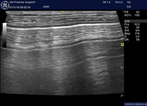
In the below clip from the Sonoscape S2 you can actually see the separation of the right ventricular free wall from the pericardium in a cat. The appearance of the hematocrit sign may be observed in hemothorax with a surface layer of anechoic fluid sitting atop a settled fine echogenic sediment. Pleural effusion is typically diagnosed by taking radiographs X-rays of the chest.
Both the trocar and the modified Seldinger techniques can be used. 81 Pulmonary pathology 82 Pleural space 83 Heart and mediastinum 84 Thoracic wall. The ultrasound US examination was performed less than 6 h after the diagnostic CT scan.
Cats presenting with pleural effusion are nearly always in respiratory distress ranging from an increased respiratory rate and effort to open mouth breathing. To characterize the effusion noting echogenicity of the fluid any loculations solid masses and pleural disease. Within 458 - 287 h after the CT scan all patients were re-examined with US in the ICU.
Lung Ultrasound Flooding In Fulminant Pulmonary Oedema In Cats And A Comparison With Pneumonia Vet Practice Support
2

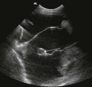
Cat Of Figure 1 Thoracic Ultrasound Revealed A Mild Hypoechoic Download Scientific Diagram

Differentiating Pericardial From Pleural Effusion Animal Ultrasound Association

Pleural Effusion And Nt Probnp In Cats

Thoracic Ultrasound

Sonography Assessment Overview Of Afast And Tfast Today S Veterinary Practice

Fig Ure 2 Abdominal Ultrasound Images Of A 6 Year Old Cat Case 3 With Download Scientific Diagram

Differentiating Pericardial From Pleural Effusion Animal Ultrasound Association
Sonography Assessment Overview Of Afast And Tfast Today S Veterinary Practice

Search Imv Imaging

Fig Ure 3 Abdominal Ultrasound Images Of A 9 Year Old Cat Case 4 With Download Scientific Diagram
Veterinary Echocardiography Newsletter 1 Effusions Animal Ultrasound Association
Spontaneous Cholecystopleural Fistula Leading To Biliothorax And Sepsis In A Cat

Feline Infectious Peritonitis

Veterinary Echocardiography Newsletter 1 Effusions Animal Ultrasound Association

Pdf Thoracic Ultrasound A Method For The Work Up In Dogs And Cats With Acute Dyspnea Semantic Scholar
Pleura Veterian Key

How To Ultrasound Detection Of Pleural Fluid Case Study Video Youtube